
Sonogram vs Ultrasound: Key Differences and How to Choose the Right One


GetScannedToday
The terms ultrasound and sonogram are often used interchangeably, but they’re not quite the same.
An ultrasound refers to the imaging technique — it uses high-frequency sound waves to look inside the body.
A sonogram is the image produced during an ultrasound scan.
In simple terms:
Ultrasound = the process
Sonogram = the image
How Do Sonograms and Ultrasounds Work?
Ultrasound machines send sound waves into the body using a handheld probe. These waves bounce off internal structures like organs, blood vessels, or a foetus. The returning echoes are captured and turned into live images: sonograms, on a screen.
There’s no radiation, making it a safer option for many clinical settings, including pregnancy.
Types of Ultrasound and Sonography: Choosing the Right Imaging Type
There are several types of ultrasound, depending on the part of the body being examined:
- Abdominal ultrasound – often used for the liver, kidneys, gallbladder, or pancreas.
- Pelvic ultrasound – used for the uterus, ovaries, bladder, and prostate.
- Obstetric ultrasound – used during pregnancy.
- Vascular ultrasound – looks at blood flow in arteries and veins.
- Echocardiogram – focused on the heart.
The type of scan chosen depends on your symptoms and what your doctor is investigating.
Sonogram vs Ultrasound vs Other Imaging Techniques: When to Choose Each
Ultrasound is ideal for soft tissues and real-time imaging: like checking a baby’s heartbeat or blood flow.
However, it has limitations.
- MRI is better for detailed images of soft tissues, like the brain or joints.
- CT scans give detailed cross-sectional images and are better for bone injuries, lung problems, or detecting cancers.
Your doctor will choose the most appropriate scan based on what needs to be seen and how urgently.
Medical Applications of Ultrasound: What Will an Ultrasound Show?
Ultrasound can help detect a wide range of conditions, such as:
- Gallstones or kidney stones
- Liver or pancreatic problems
- Heart function issues
- Problems in the womb or ovaries
- Fluid build-up or swelling
- Vascular conditions
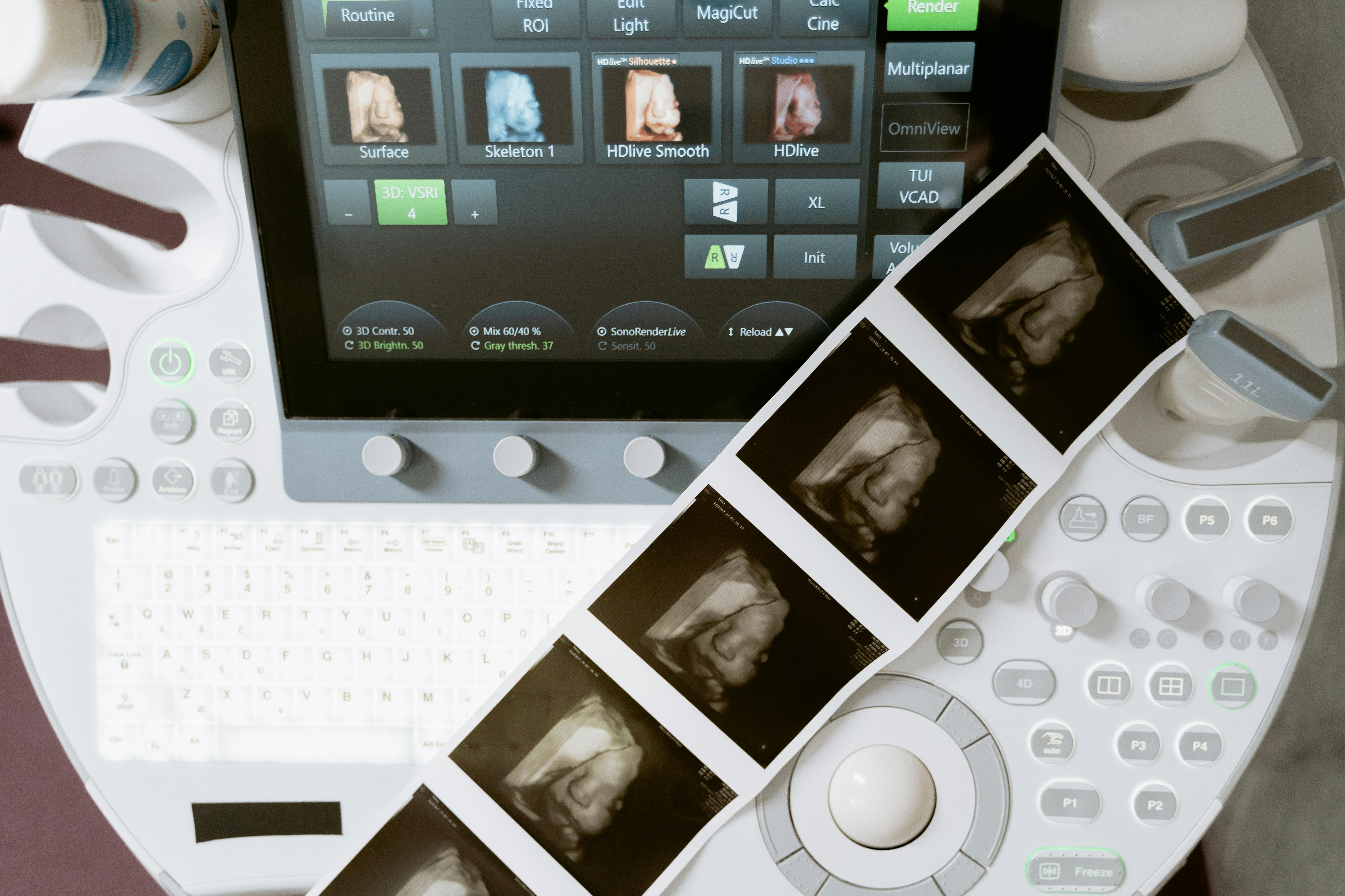
Understanding Sonography Images: What Does a Sonogram Show?
A sonogram shows a real-time image of what’s happening inside the body.
It might look like a blurry black-and-white image to most, but trained professionals can interpret it to spot abnormalities, measure structures, or check for blockages. In pregnancy, it shows the fetus moving, growing, and developing.
Safety of Sonograms and Ultrasounds: Are They Safe?
Yes, ultrasounds are generally very safe.
They use sound waves and have been widely used for decades. There’s no evidence that ultrasounds cause harm when used appropriately. That’s why they’re routinely used in antenatal care and for many other conditions.
Cost Considerations: How Much Does a Sonogram Cost?
In the NHS, most ultrasound scans are free if referred by a GP or hospital doctor.
Privately, costs vary. A basic scan can start around £50–£100 for limited checks, but more detailed or specialist scans can cost £250 or more. Prices vary depending on the clinic, location, and type of scan.
The Evolution of Sonograms: When Was the Sonogram Invented?
Ultrasound technology dates back to the 1950s. It was first used medically in Scotland, with Professor Ian Donald pioneering its use in obstetrics. Since then, it has evolved into a widely accessible and advanced imaging tool used all over the world.
Conclusion
Understanding the difference between a sonogram and an ultrasound empowers you to make informed decisions about your health. Whether you’re monitoring a pregnancy, checking for organ health, or investigating symptoms, ultrasound technology offers a safe, non-invasive way to see inside the body. Knowing when to choose an ultrasound—and understanding the images it produces—can help you navigate medical care with more confidence.
Need a private ultrasound scan in the UK?
Book your appointment today at GetScanned for fast, affordable, and expert-led diagnostic imaging—no long NHS wait times.
FAQs
1.What is the main difference between an ultrasound and a sonogram?
An ultrasound is the imaging procedure; a sonogram is the image produced.
2.Is a sonogram only used for pregnancy imaging?
No, sonograms are used to investigate many conditions affecting the abdomen, pelvis, heart, blood vessels, and more.
3.When should I choose an ultrasound over a sonogram?
You don’t choose between the two — they go hand-in-hand. Ultrasound is the scan, and a sonogram is the image it produces.
4.What types of conditions are best diagnosed with ultrasound?
Ultrasound is best for soft tissue problems — like gallstones, cysts, blood flow issues, and pregnancy monitoring.
5.Do sonograms provide the same level of detail as other imaging types like MRI or CT scans?
Not always. Ultrasound is excellent for real-time, soft tissue imaging, but MRI and CT offer more detail in certain areas like the brain, bones, or lungs.
Take a look at our news and articles


Frequently asked questions
Do I need a GP-referral?
No prior GP-referral is required. Booking with us includes a GP phone consultation and referral. Shortly after booking you will be contacted by a GP from our team who will discuss your scan and provide a referral.
How long is an MRI scan?
MRI scans generally take a bit longer than other types of scans. Individual scans take 10-30 minutes depending on the body part being scanned, overall it can take anywhere from 15 minutes to 90 minutes. You do have to lay very still for an MRI and if there is movement the scan may need to be repeated which can add some additional time.
What’s included in my booking?
With your GetScanned booking, you will receive:
- A pre-scan phone consultation with a member of our medical team.
- A referral for the scan.
- Scheduling of a private MRI scan at your preferred scanning centre.
- Access to your written report by a radiologist.
- Access to your scan images (online and downloadable).
How much is a private MRI scan?
A private MRI scan cost varies depending on the part of the body being scanned and the location the scan is performed. Generally, a private MRI scan in the UK starts at around £350, and includes the scheduling, scan itself and results.
What’s the difference between an open and closed MRI?
Closed MRI machines are the traditional and first type of MRI. They are used more frequently because they provide higher quality images, however they aren’t ideal for certain types of scan or when the patient has limited mobility. Open or wide-bore MRI machines don’t involve lying in a tight cylinder, instead they have wider openings with more space and are therefore considered better if you suffer from claustrophobia. A standing MRI or upright MRI is a new type of open MRI that allows the patient to be in various different positions, including weight bearing positions. If you would prefer an open MRI please filter by MRI type to find an open MRI near you, but please be aware it is only available at certain locations.

Still have questions?
Can't see an answer to your question? Our friendly customer care team are happy to help.
