


No items found.
What do lesions on a brain MRI mean?
GetScannedToday
Thank you! Your submission has been received!
Oops! Something went wrong while submitting the form.
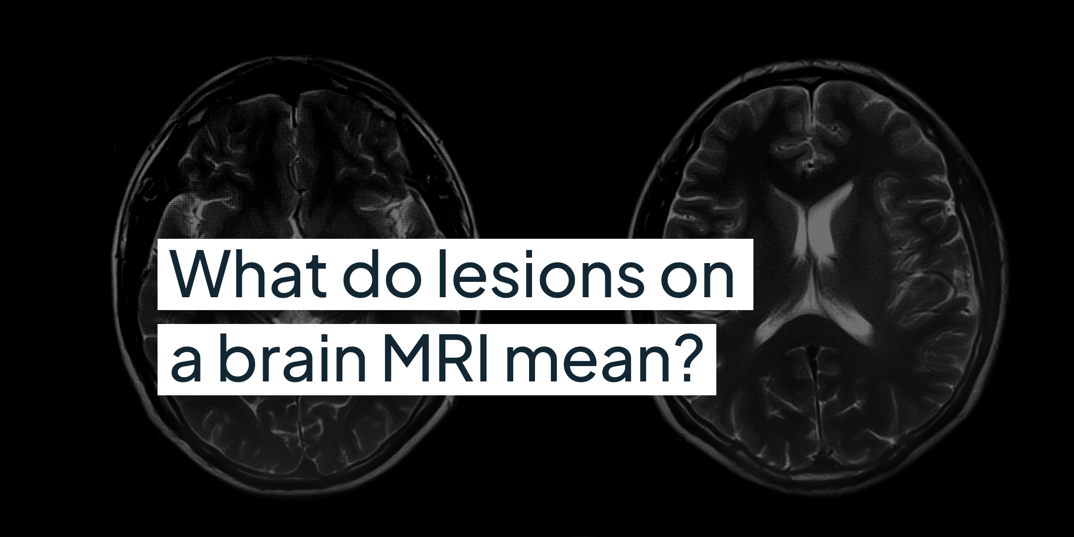
A lesion on a brain MRI is simply an area that looks different from the tissue around it. The word describes how something looks on the scan. It is not a diagnosis, and on its own it does not tell you whether something is serious.
Why the report uses this word
Radiologists use "lesion" because it describes what they can see without assuming the cause. Reports may also use related terms such as white matter hyperintensities, white spots or foci, small vessel or microvascular changes, or gliosis (a type of scarring).
The most common finding is small white spots in the brain's white matter, the wiring that connects different areas. These become more common with age, a bit like grey hair or wrinkles, and are often linked to high blood pressure, smoking, diabetes and high cholesterol. Less often, lesions are caused by multiple sclerosis (MS), small strokes that caused no symptoms at the time, migraine, inflammation or infection. Rarely, a lesion is a growth such as a tumour. Findings are common: around one in ten brain scans shows something, and most are harmless variations.
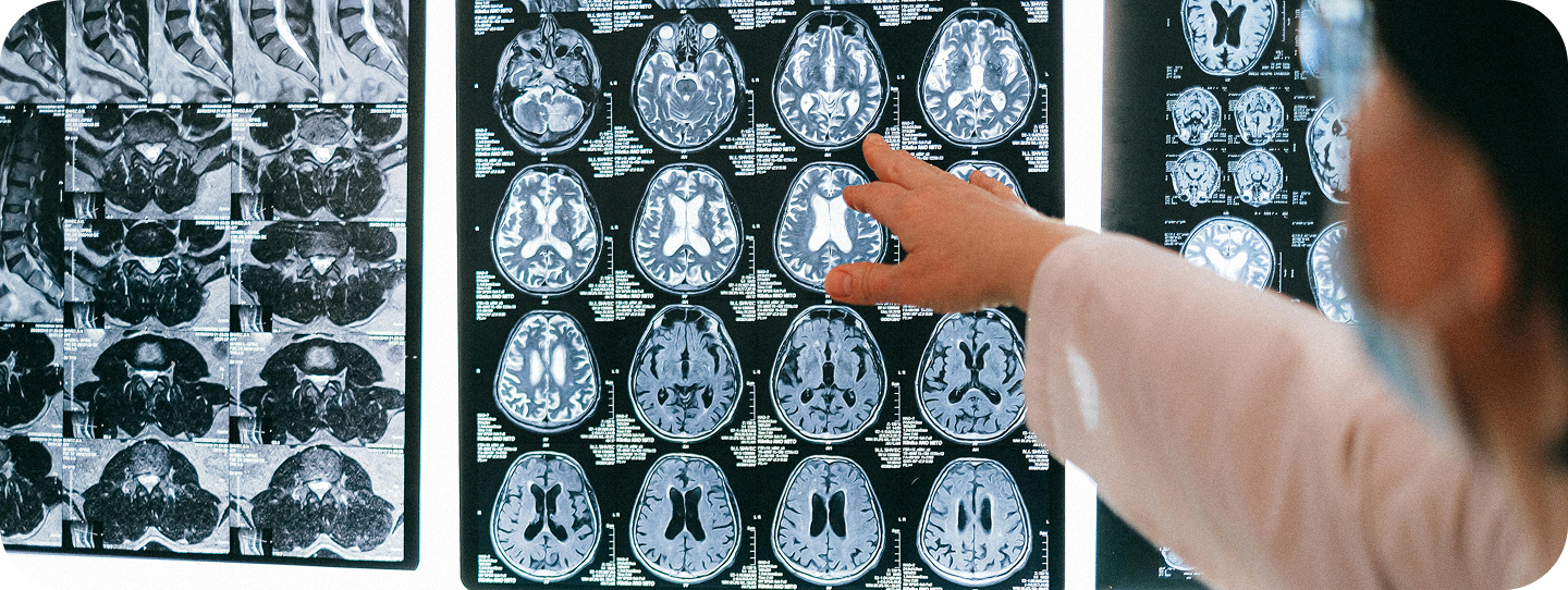
To tell these apart, a radiologist looks at the size, shape, location and pattern of any lesions. Sometimes a more detailed scan is needed, such as one using contrast dye or a scan of the spine. This is why the summary at the end of your report, often called the impression or conclusion, matters more than any single word in it.
What happens next
The doctor reviewing your results, whether your CoreVitals clinician or your GP, will consider the report alongside your symptoms and medical history. Often no further scans are needed. For age related white matter changes, the most useful next step is usually checking and managing blood pressure, cholesterol, blood sugar and smoking. If there is an open question, your doctor may suggest a follow up scan, a comparison with older images or a specialist opinion.
If a follow up scan is suggested, the type of scan matters. Some questions need contrast dye or imaging of the spine, which a standard scan may not include. If your doctor recommends further imaging, you can find out more about our private MRI scan and speak to our team about whether it suits your situation.
Book a GP appointment soon if you notice new symptoms that last or get worse, such as numbness or tingling, blurred or painful vision in one eye, problems with balance, or changes in memory.
When the situation is urgent
Call 999 if you or someone else suddenly has any of these symptoms, even if they go away:
- Face drooping on one side, arm weakness, or difficulty speaking.
- Sudden loss or blurring of vision.
- Sudden numbness or weakness on one side of the body.
- Sudden confusion, dizziness or loss of balance.
- A sudden, very severe headache.
- A first seizure.
A private MRI is never the right route in an emergency.
Take a look at our news and articles
Browse all articles


Frequently asked questions
Do I need a GP-referral?
No prior GP-referral is required. Booking with us includes a GP phone consultation and referral. Shortly after booking you will be contacted by a GP from our team who will discuss your scan and provide a referral.
How long is an MRI scan?
MRI scans generally take a bit longer than other types of scans. Individual scans take 10-30 minutes depending on the body part being scanned, overall it can take anywhere from 15 minutes to 90 minutes. You do have to lay very still for an MRI and if there is movement the scan may need to be repeated which can add some additional time.
What’s included in my booking?
With your GetScanned booking, you will receive:
- A pre-scan phone consultation with a member of our medical team.
- A referral for the scan.
- Scheduling of a private MRI scan at your preferred scanning centre.
- Access to your written report by a radiologist.
- Access to your scan images (online and downloadable).
How much is a private MRI scan?
A private MRI scan cost varies depending on the part of the body being scanned and the location the scan is performed. Generally, a private MRI scan in the UK starts at around £350, and includes the scheduling, scan itself and results.
What’s the difference between an open and closed MRI?
Closed MRI machines are the traditional and first type of MRI. They are used more frequently because they provide higher quality images, however they aren’t ideal for certain types of scan or when the patient has limited mobility. Open or wide-bore MRI machines don’t involve lying in a tight cylinder, instead they have wider openings with more space and are therefore considered better if you suffer from claustrophobia. A standing MRI or upright MRI is a new type of open MRI that allows the patient to be in various different positions, including weight bearing positions. If you would prefer an open MRI please filter by MRI type to find an open MRI near you, but please be aware it is only available at certain locations.

Still have questions?
Can't see an answer to your question? Our friendly customer care team are happy to help.
Contact Us
